Cytokinesis and karyokinesis are significant steps in the cell cycle. The term ‘Cyto’ refers to the cytoplasm, and ‘kinesis’ refers to the movement. Cytokinesis is the process involving the division of the cytoplasm of a cell. On the other side, the word ‘Karyon‘ means nucleus, and ‘kinesis’ is movement. Thus, karyokinesis depicts the nuclear division or the segregation of nuclear material.
Cytokinesis is comparatively a simpler and single-step process. In comparison, karyokinesis involves a train of events that make it complex.
Cytokinesis is the terminal step in the cell division process. So, it takes place after karyokinesis finishes. For this reason, Cytokinesis becomes dependent on karyokinesis.
In contrast, karyokinesis is the initial step in cell division. Thus it doesn’t rely on any other process and can occur independently. It may or may not be followed by Cytokinesis as per the cellular requirements.
The process of cytokinesis results in two daughter cells. It is the equal division of the cytoplasm and cell organelles of the parent cell. Whereas the karyokinesis generates two daughter nuclei by distributing nuclear material equally.
In this context, we aim to illustrate major differences between cytokinesis and karyokinesis. We will also be studying their mechanism, comparison chart, stages with diagrammatic representation.
Content: Cytokinesis Vs Karyokinesis
Comparison Chart
| Basis of Comparison | Cytokinesis | Karyokinesis |
|---|---|---|
| Meaning | Cyto stands for cell and kinesis for movement. Thus, it is the process where the cell divides causing the division cytoplasm and organelles to form new cells. | Karyo means cellular nucleus and kinesis is movement (division). It is the process of division of nucleus and its components. |
| Division | Protoplasm of the parent cell divides into two daughter cells | During this, the parent nucleus divides to generate two daughter nuclei. |
| Function | Dividing the cytoplasm and cellular organelles of parent cell into two daughter cells. | Dividing nuclear material of parent nucleus to form two daughter nuclei. |
| Dependency | It can only take place after karyokinesis and thus is dependent on it. | It is independent of Cytokinesis i.e. it may or may not be followed by the Cytokinesis. |
| Occurrence | Occurs at the end of M-phase after the karyokinesis is completed. | Occurs during the initial stage of M-phase before Cytokinesis. |
| During cell division | It is the terminal step of cell division. | It is the beginning step of cell division. |
| Distribution | Distributes equal amount of cytoplasm and other cellular components equally among the two resultant daughter cells. | Distributes the nuclear material equally into the daughter nuclei. |
| Complexity | Quite simpler process. | Intricate process involving the segregation of nuclear components. |
| Constriction | Cell membrane generates a constriction or cleavage furrow that segregates the parent cell into two new cells. | Nuclear membrane develops a constriction i.e. dumbbell shaped. |
| Events | • Formation of cleavage furrow. • Development of cell plate at the centre of division. | • Formation of Spindle fibres. • Movement of chromosomes. |
| Types of Division | Two kinds of division are seen during Cytokinesis: - 1. Symmetric 2. Asymmetric | There are no types karyokinesis. |
| Stages | There are four stages: 1. Initiation 2. Contraction 3. Membrane Insertion 4. Termination | There are four stages: 1. Prophase 2. Metaphase 3. Anaphase 4. Telophase |
What is Cytokinesis?
Cytokinesis is a simple process but a crucial process in the cell cycle. This step gives birth to two new daughter cells from the parent cell. The new cells will contain an equal amount of cellular components.
We can’t consider cell division as complete until cellular components are physically apportioned. This makes cytokinesis much essential.
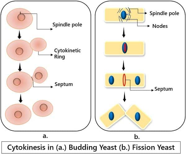
In almost all organisms, cytokinesis is the last stage of cell division. And it may differ from cell to cell and from organism to organism.
Characteristic of Cytokinesis
- It is the final step of cell division which terminates the cell cycle by dividing the parent cell into two.
- It occurs as the terminal step in both sorts of division-meiosis and mitosis. The process begins with the formation of the contractile ring. This ring is made up of cytoskeletal proteins.
- During the process, the cell membrane gets fused initially. Later, it breaks as the ring contracts.
- The division leads to the equal distribution of the entire protoplasm. For example, cytoplasm, nucleus as well as other organelles.
- In some eukaryotes like plants, cell plate formation takes place. This cell plate matures to generate a cell wall.
- While in animals, only the cleavage furrow is sufficient for the segregation of a parent cell into two daughter cells.
Types of Cytokinesis
Cytokinesis is mainly of two kinds:
a. Symmetrical
Here, the parent cell divides into two daughter cells with similar shapes, sizes and amounts of cellular content.
Example: Spermatogenesis.
b. Asymmetrical
It is the division where the parent cell divides and forms two unequal daughter cells.
Example: Oogenesis.
Mechanism of Cytokinesis
In Animals
- The mechanism of cytokinesis varies as per the cellular organisation of the organism.
- In animals, cells lack cell walls. Thus the process of cytokinesis occurs with the formation of the contractile ring.
- This contractile ring constitutes actin fibres which are proteins by nature.
- This ring is developed right inside the cell membrane at the former metaphase plate.
- The actin filaments form a fissure by pulling the equator of the cell inwards.
- The fissure or crack will eventually give rise to the cleavage furrow.
- The furrow intensifies and gets deepened with the gradual contraction of the actin ring.
- As the cleavage reaches to its edge, the membrane separates, giving rise to two new daughter cells.
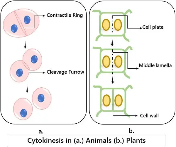
In Plants
- The plant cells contain a cell wall, due to which the daughter cells must also possess it.
- Because of this, Golgi bodies of the dividing cell start gathering various materials. Such as structural proteins, essential enzymes and lots of glucose.
- Further, they break into vesicles and scatter throughout the parent cell.
- In plants, cytokinesis takes place with the formation of the cell plate.
- The cell plate is generated in the middle portion of the parent cell with the help of vesicles and microtubules.
- The vesicles containing proteins, carbohydrates, lipids, enzymes etc. are accumulated in the mid-region of the Phragmoplast microtubule array extending from the centre towards the cell wall. This structure is known as a cell plate.
- Later, the cell wall components like cellulose, hemicellulose, pectic etc., start to traffic around this cell plate, leading to its maturation. The cell plate stretches until it fuses the peripheral cell wall.
What is Karyokinesis?
Karyo stands for nucleus while kinesis depicts the movement i.e., the division here. Karyokinesis is just as essential as Cytokinesis in the cell cycle.
Definition of Karyokinesis
We can define karyokinesis as:
“The process where the nucleus of the parent cell is divided into two daughter nuclei”.
Nuclear material synthesised during the S-phase gets converted into chromosomal fragments. These genetic fragments are equally distributed among the daughter nuclei. At the time of division, karyokinesis occurs in the initial phase. Thus, cytokinesis occurs only after the completion of karyokinesis.
Every cell of the living body has to rejuvenate and regenerate while dividing. Karyokinesis reassures the passage and equal distribution of hereditary material while the cell is dividing. Thus, it is a crucial step in cell division.
Characteristics of Karyokinesis
- It is the first step in the cell division process.
- The process splits and distributes the nuclear material between the two daughter nuclei.
- The nuclear envelope, i.e., the nuclear membrane completely dissociates at the beginning of karyokinesis. Later this envelope appears back as the process ends.
- At this stage, the replicated nuclear material appears as the chromosomes.
- Nuclear material continuous changes its configuration from chromatin to chromosome throughout the process.
- In the end, the chromosomes get decondensed again to form the chromatid material.
Mechanism of Karyokinesis
The process of the cell cycle initiates at interphases. During interphase, the cell prepares itself for division, and the nuclear material replicates. This material is in condensed form with a thick thread-like appearance. Also, interphase synthesises all the essential proteins for karyokinesis, including centromere and kinetochores.
During prophase, chromatid threads condense into short fragments of chromosomes. The metaphase aligns these chromosomes at the centre. In anaphase, sister chromatids segregate as two daughter chromosomes from the centromere.
Later, telophase leads to the separation of nuclear material between two daughter cells. The nuclear membrane arises again to produce a membrane-bound nucleus.
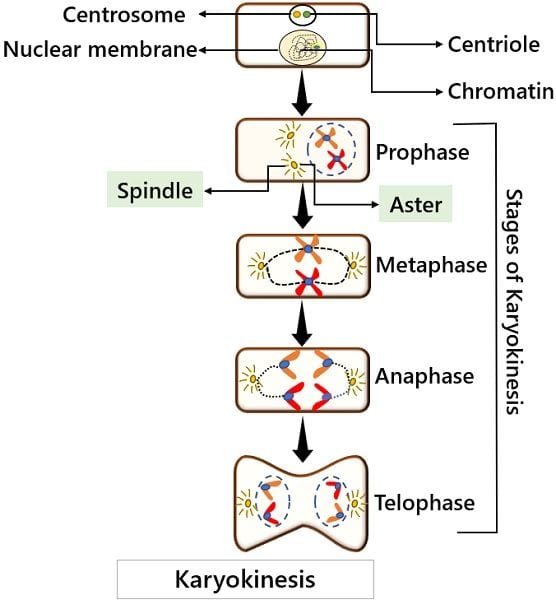
Stages in Karyokinesis
The process of karyokinesis occurs in four consecutive phases. These are as follows:
-
Prophase
- It is the first phase that initiates the process of karyokinesis.
- During this phase, the nuclear membrane dissolves and fragments into small vesicles.
- All the membrane-bound organelles like mitochondria, ER, and Golgi bodies dissociate. Further, they move at the peripheral edge of the cell.
- The nucleolus also disappears.
- Whereas the centromeres disperse towards the opposite poles of the cell.
- Microtubules outspread between these centromeres. The microtubule fibres elongate here. This elongation extends the centromeres at their maximum distance.
- Most importantly, the chromatin material gets condensed to form chromatid threads.
- The sister chromatids start coiling with the help of condensin proteins.
-
Metaphase
The metaphase has two sub-phases: prometaphase and late metaphase.
a. Prometaphase
- It is the first change phase.
- The processes started during the prophase continue during this phase.
- The remaining fragments of the nuclear membrane dissociate.
- The microtubules start forming more and more spindles. The spindles further align and assemble across the dimensions of the prior nuclear zone.
- The chromosomes are now completely formed with distinctive condensed structures.
- A protein named kinetochore attaches the sister chromatids together at the centromeric region.
- This kinetochore is responsible for attracting and attaching the chromosomes to the spindles.
b. Late Metaphase
- This is the later change phase.
- While this phase, the chromosomes align themselves in the equatorial region, i.e., in the midway of the cell.
- This alignment leads to the formation of a metaphase plate.
- Here, the chromosomes are at their maximum condensed state.
-
Anaphase
- It is the upward phase where the cohesion proteins degrade. This leads to the segregation of the sister chromatids from the centromere.
- As the spindle contracts, it pulls the centromere along with the chromosome. In this way, both the chromosomal threads move in opposite directions.
- Later, the cell elongates and takes a flattened oval shape.
- This is because the polar microtubules slide at the equatorial region at the place of overlapping.
-
Telophase
- It is the distant phase. Here the chromosomes reach at the proximal end or periphery of the cell.
- At the opposite ends, the chromosomes again start to decondensed. This process unravels the chromosomes to regain their chromatin configuration.
- Mitotic spindles degenerate into tubulin fibres through depolymerisation.
Note: These tubulin monomers aid each daughter cell in assembling cytoskeletal components.
- The nuclear membrane envelops the decondensed chromatid material. This gives rise to two new nuclei at both ends.
- The nucleosomes restore back.
Key Difference Between Cytokinesis and Karyokinesis
- Cytokinesis is the division of cells and their components. Whereas karyokinesis refer to the division of nuclear and nuclear material.
- The protoplasm of the parent cell divides into two daughter cells during Cytokinesis. While the karyokinesis gives rise to two daughter nuclei
- Cytokinesis is a dependent process. It occurs after the termination of karyokinesis. But the karyokinesis is an independent phase. This is because it doesn’t have to rely on any other process.
- Cytokinesis occurs as the terminal step for cell division. In contrast, karyokinesis is the initial step that appears at the beginning.
- Cytokinesis distributes the cytoplasmic components among the daughter cells. Contrarily, the karyokinesis equally distributes the chromosomal material between two daughter nuclei.
- Cytokinesis is a simpler process than that of karyokinesis. This is because karyokinesis has its four typical stages.
Summary
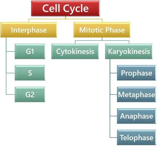
The cell cycle comprises many crucial stages. Each has its own purpose. The process of cell division initiates at the interphase. Later during the M-phase, the actual and the visible division occurs.
A parent cell to get divided into two new daughter cells, two processes are most important.
They are karyokinesis and Cytokinesis. Karyokinesis segregates the nuclear material between two new daughter nuclei. After the formation of two nuclei, the parent cell elongates and separates into two daughter cells. This process of formation of two daughter cells is Cytokinesis.
Leave a Reply